
Skin Cancer Near the Eye: Signs & Treatment
Dr Varun Harish, FRACS Plastic Surgeon
First published:
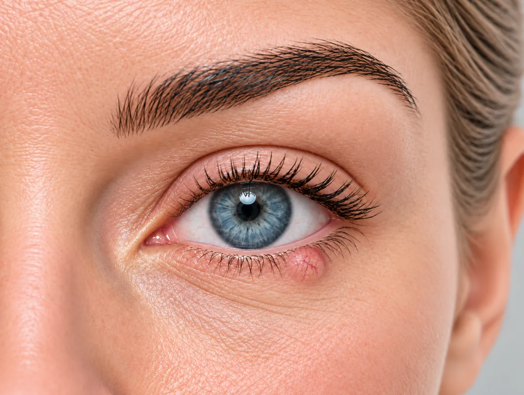
Skin cancer near the eye needs careful treatment because the eyelids help protect the eye, support normal blinking and keep the eye surface lubricated with tears. Removing the cancer is important, but the reconstruction also matters because the eyelid has important functions as well as a visible appearance.
Doctors may also use the term periocular skin cancer, meaning cancer arising in the skin around the eye. This is different from a cancer that starts within the eye itself.
Treatment usually starts with assessment and, in some cases, a biopsy to confirm the diagnosis. The skin cancer is then removed, and the wound (‘hole’ left behind after cancer removal) is repaired in a way that suits its size and location. Small wounds may be stitched up. Larger wounds, or those close to the eyelid edge or inner corner of the eye, may need more detailed eyelid reconstruction.
What “skin cancer near the eye” means
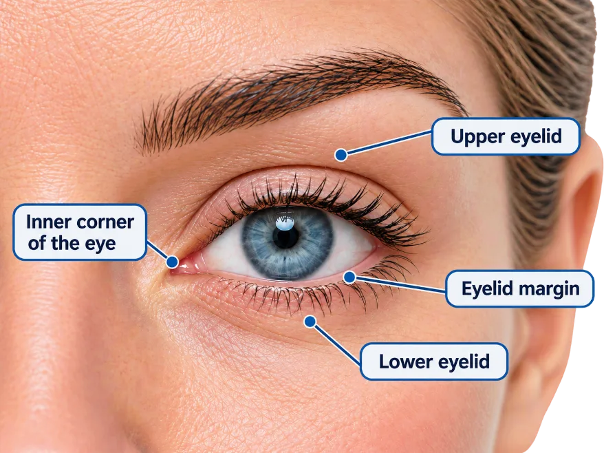
Skin cancer near the eye means a skin cancer arising on the eyelid or the nearby facial skin. This can include the upper or lower eyelid, skin under the eye, the inner or outer corner of the eye, and the eyebrow. Treatment needs to achieve adequate cancer clearance while preserving or reconstructing tissues needed for normal eye closure, eyelid position, corneal protection and tear drainage.
It is useful to think of treatment as having two connected parts. First, the cancer needs to be removed with an appropriate margin or margin-checking process. Second, the wound (‘hole’ left behind after cancer removal) needs to be repaired.
The repair depends on the final wound. Some wounds can be closed with simple stitches. Others may need nearby skin to be moved into the area (a ‘flap’), a piece of skin transplanted from elsewhere (a ‘skin graft’), or a more complex eyelid repair.
The most important factors are the cancer type, the exact location, whether the eyelid edge or tear duct area is involved, how the margins are checked and what repair will best protect eye comfort.
Skin cancer near the eye: At a glance
| Warning signs | A persistent or growing lump, non-healing sore, repeated bleeding or crusting, pearly or shiny skin, local eyelash loss or distortion of the eyelid |
| Diagnosis | Suspicious lesions are examined and usually require biopsy |
| Cancer removal | Treatment may involve standard excision, margin-controlled excision with frozen-section assessment, or Mohs surgery depending on the tumour and its location |
| Reconstruction | The final hole after cancer removal may be stitched up (closed) or require a form of reconstruction |
| Why location matters | Different areas around the eye have different functional demands, including eyelid support, eye closure, lash-line alignment and tear drainage |
Why skin cancer around the eye is different
The eyelids are not just skin. They have important functions. The eyelids protect the eye surface (‘cornea’), spread tears to lubricate the eye surface, support normal blinking and help the eye close. The upper eyelid also needs to lift clearly to allow the eye to see and to close fully for eye protection. The inner corner of the eye is close to the tear drainage system.
Reconstruction around the eye is therefore not simply about closing a wound. A small change in eyelid position after skin cancer removal near the eye can alter any of the above, and may cause irritation, watering, dryness or exposure of the eye surface. Reconstruction is about restoring a stable eyelid, protecting the eye surface and placing scars in a way that respects the natural folds of the face.
What does skin cancer near the eye look like?
Skin cancer around the eye does not always look dramatic.
Some cancers form an obvious growing lump or raw area (ulcer). Others may resemble a small area of dry skin, a persistent red patch, a sore that does not resolve, a blocked gland, cyst or an area that repeatedly crusts and then seems to improve.
Features that deserve assessment include:
- a sore that does not heal
- repeated crusting or bleeding
- a lump that progressively enlarges
- a pearly or shiny spot, sometimes with visible fine blood vessels
- persistent redness, scaling or thickening
- missing eyelashes
- distortion of the eyelid edge
- pain, tenderness or altered sensation
- a lesion that returns after previous treatment
- new or changing pigmentation
None of these features proves that a lesion is cancer. Benign conditions can look similar.
The more useful warning signs are often persistence, recurrence and progressive change rather than appearance alone.
Quick answer — Signs that deserve assessment
Skin cancer near the eye may appear as a persistent lump, pearly or shiny spot, red or scaly patch, non-healing sore, or an area that repeatedly crusts or bleeds. Local eyelash loss, ulceration, progressive enlargement or distortion of the eyelid can also be concerning. These findings are not diagnostic by themselves, but a persistent or changing lesion should be assessed.
Types of skin cancer that occur around the eye
The main skin cancers that occur around the eye are basal cell carcinoma, squamous cell carcinoma and melanoma.
Basal cell carcinoma — BCC
Basal cell carcinoma is the most common skin cancer around the eye.
An eyelid BCC may appear as a slowly enlarging lump, pearly or shiny area, persistent ulcer or sore, or an area that repeatedly bleeds and crusts. Fine surface blood vessels may sometimes be visible.
BCC often grows slowly, but if left untreated it can progressively damage nearby tissues. A larger cancer also creates a larger reconstructive problem after removal.
Squamous cell carcinoma — SCC
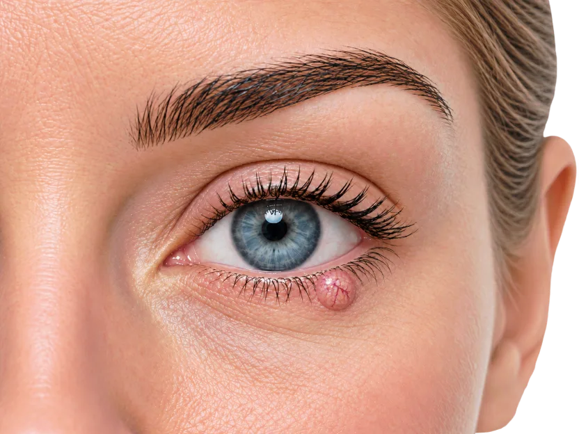
Squamous cell carcinoma is less common around the eye than BCC but can behave more aggressively.
An SCC may appear as an enlarging scaly or crusted lesion, firm lump, a sore or pimple. It may grow more quickly, bleed or become tender.
Some SCCs have the ability to spread to lymph nodes or elsewhere. However, the risk is not the same for every SCC.
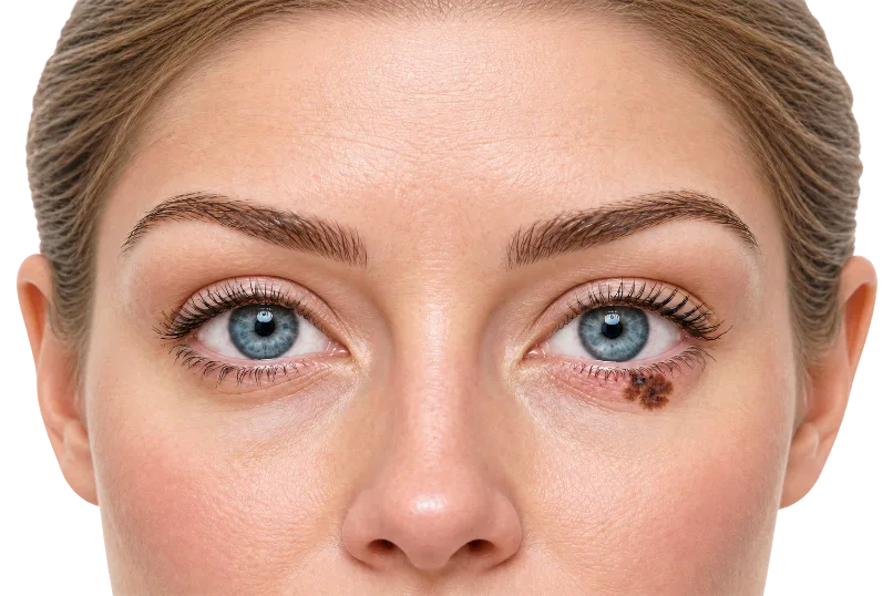
Melanoma
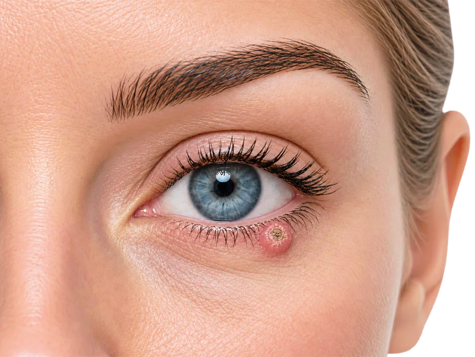
Melanoma involving eyelid skin is uncommon but important. It may present as a new or changing pigmented (mole-like) lesion, although melanoma is not always darkly coloured.
Melanoma requires its own treatment pathway. Decisions about surgery and whether additional tests are required depend on the features of the melanoma.
Rarer cancers
Sebaceous carcinoma and Merkel cell carcinoma are uncommon but can behave differently from BCC, SCC or melanoma. Sebaceous carcinoma is particularly important because it can sometimes resemble a stye. These cancers may require specialised or multidisciplinary management.
Research and guideline relevance
Dr Varun Harish was first author of peer-reviewed research examining surgical margins and prognostic factors in eyelid melanomas. The study reported what was then the largest published series of its kind. This research is cited in Australian clinical practice guidelines for melanoma, including evidence considered in relation to excision margins for eyelid melanoma.
The exact treatment depends on the diagnosis. A spot near the eye should not be treated as a minor cosmetic blemish if it is growing, bleeding, crusting, changing, painful or distorting the eyelid.
When to seek urgent review
Seek urgent medical attention if a lesion around the eye is growing quickly, bleeding repeatedly, causing pain, changing vision, preventing the eyelid from closing, pulling the eyelid out of position, or causing increasing redness, swelling or discharge.
A new or changing lesion near the eye should be assessed by a GP, dermatologist, ophthalmologist, plastic surgeon or other appropriately trained clinician. The right pathway depends on the appearance, location and suspected diagnosis.

How skin cancer near the eye is diagnosed
Assessment starts with examining the lesion and surrounding tissues.
Clinical examination, dermoscopy or photographs may help determine how suspicious a lesion appears. However, appearance does not reliably establish the exact diagnosis.
When tissue confirmation is required, the diagnosis is established by histopathological examination of tissue obtained by biopsy or excision. The pathologist can determine whether the lesion is cancer and identify the tumour type and relevant microscopic characteristics.
Not every lesion necessarily requires a separate biopsy procedure before it is removed.
Quick answer — How is skin cancer near the eye diagnosed?
A lesion may look suspicious clinically, but appearance alone cannot reliably determine whether it is cancer or what type of cancer it is. The diagnosis is generally confirmed by sampling the suspicious area by biopsy or removal.
Why location around the eye matters
The eyelids are not simply pieces of skin.
They have several important functions:
- protect the surface of the eye
- spread tears across the eye surface
- allow normal blinking
- close to protect the eye
- maintain contact with the eye surface
- contribute to normal tear drainage
The upper eyelid also needs to move adequately to open the eyes and then close fully again.
The inner corner of the eye is closely related to the tear drainage system.
This is why even small skin cancers in this area can create a significant functional problem around the eye.
How different locations change the problem
| Location | Main concern | Why reconstruction planning matters |
|---|---|---|
| Upper eyelid | Opening and closing the eye | Repair must avoid tightness that limits eyelid movement or affects vision |
| Lower eyelid | Lower-lid support | Repair must avoid pulling the lid down, which can cause watering, dryness or exposure |
| Eyelid margin | Lash line and lid edge alignment | Poor alignment can cause notching, irritation, asymmetry or rubbing on the eye |
| Inner corner of the eye | Tear drainage | Consideration for tear drainage pathways after surgery |
How skin cancer near the eye is removed
Surgery is a common treatment for skin cancer around the eye, but there is no single operation suitable for every tumour.
The removal method depends on factors including:
- the cancer type
- tumour size and depth
- exact location
- whether the borders are clearly defined
- whether the cancer has been treated before or recurred
- how much useful normal tissue surrounds it
The objective is adequate cancer removal while preserving as much uninvolved functional tissue as is safely possible.
Standard surgical excision
Some skin cancers near the eye can be treated with conventional surgical excision. The tumour is removed with an appropriate margin of surrounding tissue and sent to pathology. The pathology report then helps determine whether the margins are clear and the tumour has been completely removed.
Depending on the cancer and treatment pathway, reconstruction may occur during the same procedure or may be delayed until satisfactory clearance has been established.
Margin-controlled excision
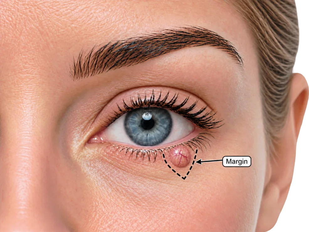
Margin-controlled surgery allows more detailed assessment of the edges of removed tissue. Around the eye, this can be valuable because relatively small amounts of preserved healthy tissue may make reconstruction easier and help retain function. Different methods of margin control are used. These may include frozen-section assessment during surgery.
Mohs surgery
Mohs surgery is a specialised form of margin-controlled skin cancer treatment. The cancer is removed in stages and the tissue is examined microscopically. If tumour remains at a particular edge, additional tissue can be removed from that area. After Mohs surgery, the wound may be repaired by a plastic surgeon.
Quick answer — Does every skin cancer near the eye need Mohs surgery?
No. Mohs surgery can be useful for certain skin cancers, but it is not required for every lesion near the eye. Many cancers can be appropriately managed with standard excision or other forms of margin-controlled excision.
The two decisions: removing the cancer and repairing the wound
People often understandably think of skin cancer surgery as one operation. Near the eye, it is more useful to think of two linked decisions.
The first decision is how the cancer should be removed and how cancer clearance will be confirmed. This depends on the diagnosis, site, size, recurrence risk and pathology.
The second decision is how the wound should be repaired once the cancer has been removed. This depends on the final wound size, depth and location. The repair may be simple if the defect is small and away from key eyelid structures. It may be more complex if the wound involves the lower eyelid, eyelid margin, inner corner of the eye, or tear duct area.
This distinction matters because cancer removal and reconstruction may occur as part of one integrated surgical plan or as separate stages. Planning both decisions from the outset helps ensure that the method of cancer clearance and the anticipated reconstruction are considered together.
Quick answer — Cancer clearance before reconstruction
The priority for treatment of skin cancers near the eye is adequate cancer clearance. Reconstruction is then planned around the ‘hole’ or wound that remains. In some cases cancer removal, margin assessment and reconstruction occur during the same procedure; in others, definitive reconstruction is delayed until satisfactory clearance has been established.
Why the wound can be larger than the visible skin cancer
The visible skin cancer does not always show the full extent of the disease. A margin of surrounding tissue is often removed to reduce the risk of leaving cancer cells behind. If pathology shows cancer at the edge, more tissue may need to be removed.
This is why the final wound can be larger than expected. Around the eye, this can be confronting because the eyelids and surrounding facial skin are so visible. However, reconstruction is planned around the final defect after cancer removal, not just the original spot on the skin.
Surgical margins and confirming cancer clearance
The visible edge of a skin cancer does not always represent its full microscopic extent. This is why the edges, or margins, of the removed tissue are assessed.
A clear or negative margin means tumour is not seen at the examined edge. A positive margin means tumour reaches the edge and additional treatment may be required. A close margin means the tumour does not reach the edge but lies relatively near it; the significance depends on the cancer type and pathological features.
Around the eye, margin assessment is particularly important because additional tissue removal can materially change the reconstructive problem.
Learn more about why surgical margins matter in skin cancer surgery.
Who is the right specialist for skin cancer around the eye?
There is no single answer for every patient. The right specialist depends on what the cancer involves, how the margins will be checked and what kind of reconstruction is needed.
Depending on the cancer, treatment may require expertise in skin cancer surgery, margin assessment and facial reconstruction. More complex cases may also require input from specialists in eyelid, tear-drainage or eye surgery.
The important question is therefore not simply which specialty treats the cancer, but whether the treatment plan considers cancer clearance, the expected surgical defect and reconstruction together.
A plastic surgeon can often plan both the cancer removal and the reconstruction as part of the same treatment pathway. This allows the likely defect, margin strategy and reconstructive options to be considered before tissue is removed.
Reconstruction after skin cancer removal around the eye
Reconstruction near the eye is not simply about covering a hole in the skin. The most appropriate reconstruction after skin cancer surgery is therefore chosen after considering the final wound, rather than simply the cancer diagnosis.
Quick answer — Why reconstruction near the eye is different
Reconstruction after skin cancer near the eye needs to restore more than skin coverage. Depending on the defect, the repair may need to protect eye closure, eyelid position, the lash line, the cornea and tear drainage. Reconstruction may involve direct closure, a skin graft, a local flap or a more complex repair of deeper eyelid tissues.
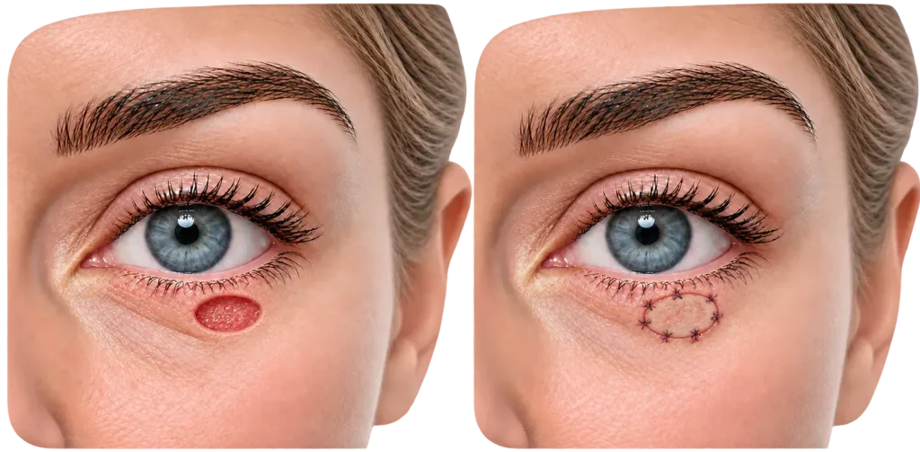
Direct closure
Direct closure means the wound edges are brought together with stitches. This may be suitable for smaller wounds where the surrounding skin has enough movement and closure will not distort the eyelid. Around the eye, direct closure is only appropriate if it can be done without pulling down the lower eyelid, distorting the lash-line or preventing comfortable eye closure.
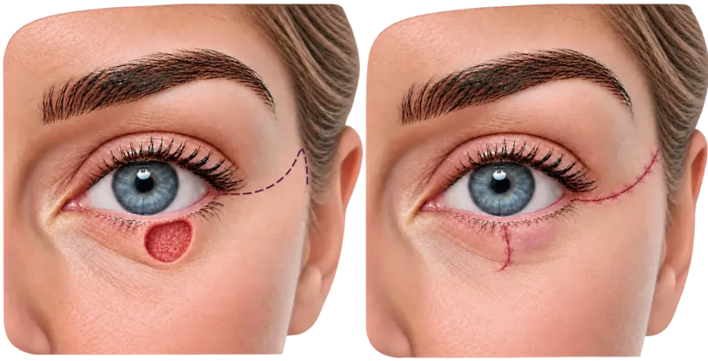
Local flap
A local flap moves nearby skin into the wound while keeping its own blood supply. Around the eye, flaps can be useful because nearby skin often provides a better match in colour, texture and thickness than skin taken from a distant site.
Skin graft
A skin graft transfers skin from another part of the body to the wound. A graft may be useful when local skin movement is limited or when a flap would create too much tension. Grafts can be helpful in selected cases, but they may differ in colour or texture and need careful follow-up because contraction during healing can affect eyelid position.
A graft may initially look different from the surrounding skin and can change during healing.
When deeper eyelid tissues are involved
Some cancers involve more than superficial skin. The eyelid contains skin and muscle externally, structural support internally and a smooth lining that moves against the eye. If a cancer involves the eyelid margin or deeper layers, reconstruction may need to restore these deeper components as well as provide skin cover.
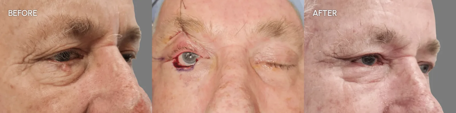
Reconstruction in stages
At times, treatment is deliberately performed in two stages.
Staging may be relevant when:
- cancer clearance is being confirmed before definitive reconstruction
- the defect is large or structurally complex
- deeper eyelid tissues need to be reconstructed
- a staged technique offers a safer way to restore support or lining
A staged repair can sound concerning, but in selected cases it is chosen because it improves control over healing, eyelid position and tissue support.
Local flap or skin graft: which is better around the eye?
Neither a local flap nor a skin graft is automatically better around the eye. The better option depends on the wound, the surrounding skin, the eyelid position and the direction of tension needed to close the defect.
A local flap uses nearby skin and soft tissue. This can be helpful on the face because nearby tissue often has a closer colour, thickness and texture match.
A skin graft uses skin transferred from another site. This can be useful when moving nearby tissue would pull the eyelid out of position or when local skin is limited. The trade-off is that grafted skin may look slightly different from surrounding eyelid skin and can contract as it heals.
For some wounds, direct closure is preferable to either a flap or a graft. For other wounds, a flap, graft or combined approach may be needed. The aim is not to choose the most complex reconstruction. The aim is to choose the repair that closes the wound safely while protecting eyelid function.
| Feature | Local flap | Skin graft |
|---|---|---|
| Where the tissue comes from | Neighbouring skin and soft tissue | Skin transferred from another body site |
| Blood supply | Remains attached to its blood supply | Develops a new blood supply from the wound beneath it |
| Colour and texture match | Often very good because tissue comes from nearby | May differ somewhat from surrounding eyelid skin |
| Effect on surrounding tissues | Moving tissue creates and redistributes tension | May avoid pulling neighbouring eyelid skin into the wound |
| Healing behaviour | Can initially appear swollen | Can change in colour and contract as it heals |
Recovery after skin cancer surgery near the eye
Swelling and bruising are common after surgery around the eye. The eyelid skin is thin, so bruising may look more dramatic than the size of the operation suggests. Temporary tightness, watering, mild irritation or blurred vision from ointment can occur.
Postoperative instructions vary depending on the repair. Some patients may be advised to use ointment on the suture line, lubricating eye drops, a dressing or protective pad, or cold compresses in the early swelling phase. Activity restrictions, wound care and when to restart usual medications should follow the treating doctor’s instructions.
Sutures may be removed at follow-up, although some stitches dissolve. Skin grafts, flaps and more complex eyelid repairs may require closer early review than a simple direct closure. Follow-up also allows the surgeon to check pathology, wound healing, eyelid position and eye comfort.
Skin grafts can initially differ in colour and texture from the surrounding skin before settling. Local flaps may initially appear swollen or bulky. Both continue to change as swelling resolves and scars mature.
The early appearance is not the final result. Scars around the eye change over months. Redness, firmness and swelling usually settle gradually. More complex reconstructions may take longer to mature, and some patients may need later adjustment if eyelid position, scar tightness or contour remains a problem.
Quick answer — How long does recovery take after eyelid skin cancer reconstruction?
Early healing usually occurs over the first few weeks, but bruising, swelling, scar firmness and colour change can take considerably longer to settle. Larger flaps, skin grafts and staged eyelid reconstructions generally take longer to mature than a small direct closure.
When assessment by a reconstructive plastic surgeon may be useful
Assessment by a reconstructive plastic surgeon may be useful when skin cancer removal near the eye is likely to require more than simple closure.
This includes lesions near the lower eyelid, eyelid margin, inner corner of the eye, cheek–eyelid junction, eyebrow, nose–cheek junction or other facial areas where eyelid position, scar placement, contour and tissue movement are important.
A plastic surgeon can assess the lesion, perform biopsy or excision where appropriate, plan how cancer clearance will be established, and reconstruct the resulting defect. Considering cancer removal and reconstruction together from the outset can be particularly useful around the eye, where the amount and direction of tissue removed can directly influence the reconstructive options available.
Some cancers may require a different or multidisciplinary pathway because of their pathology or involvement of specialised eye structures.
Questions to ask before surgery
- Bring the biopsy result if available.
- Ask whether the lesion involves the eyelid margin, lower eyelid, inner corner of the eye or tear duct area.
- Ask how margin clearance will be confirmed.
- Ask whether reconstruction is expected to occur during the same procedure or after margin confirmation.
- Ask whether deeper eyelid support or tear drainage structures may be involved.
- Ask whether repair is likely to involve direct closure, a local flap, a skin graft or a more complex eyelid reconstruction.
- Ask what symptoms after surgery should prompt urgent review.
Frequently asked questions
Is skin cancer near the eye the same as eye cancer?
No. Most cancers discussed on this page begin in the skin of the eyelid or surrounding face. They are different from cancers arising inside the eyeball or from structures on its surface.
Do I always need a biopsy before the whole lesion is removed?
No. Some lesions are biopsied first because the diagnosis will change the treatment plan. A small lesion may sometimes be completely removed by the biopsy itself. The appropriate approach depends on the suspected diagnosis, location and planned reconstruction.
Will skin cancer surgery near the eye affect my vision?
Most skin cancers around the eye arise from the surrounding skin rather than the eyeball itself, and treatment is planned to protect the eye. However, swelling, ointment or irritation can temporarily blur vision after some procedures. Larger or more advanced cancers can involve deeper structures and may require more complex multidisciplinary treatment. New reduction in vision, significant eye pain or inability to close the eye after surgery requires urgent medical assessment.
Who is the right specialist for skin cancer near the eye?
A plastic and reconstructive surgeon can assess skin cancer near the eye, perform biopsy or excision where appropriate, plan cancer clearance and reconstruct the resulting defect. This integrated approach can be particularly useful where removal of the cancer may affect eyelid position, eye closure or surrounding facial tissues. Some cancers or deeper eye-related problems may require additional specialist involvement.
Will I need a flap or skin graft?
Not always. Small wounds may be closed directly (stitched up). A flap or graft may be considered if direct closure would distort the eyelid, if the wound is too large, if local skin movement is limited or if the defect is in a functionally sensitive area.
What is the difference between a local flap and a skin graft?
A local flap moves nearby skin and soft tissue into the wound while keeping its blood supply attached. A skin graft transfers skin from another part of the body and relies on the wound bed to heal. Around the eye, a flap may offer a better tissue match, while a graft may avoid pulling nearby eyelid skin out of position. The best option depends on the hole or defect after cancer removal, eyelid tension, wound depth and surrounding tissue.
Can skin cancer surgery near the eye change the shape of my eyelid?
It can, especially if the cancer is large, close to the eyelid margin or involves the lower eyelid. The aim of reconstruction is to reduce distortion and protect eye comfort, but risks such as asymmetry, lower-lid pull-down, lid-margin irregularity or need for revision should be discussed before surgery.
About Dr Varun Harish
Dr Varun Harish is a Specialist Plastic, Reconstructive and Burns Surgeon. His practice includes skin cancer surgery and reconstruction after skin cancer removal, including facial sites where scar placement, contour, function and tissue movement are important.
Dr Harish is a Fellow of the Royal Australasian College of Surgeons in Plastic and Reconstructive Surgery and a Clinical Senior Lecturer at the University of Sydney. His public hospital work includes reconstructive surgery and complex wound management.
Dr Harish has specific academic experience in eyelid melanoma. Dr Harish completed a Master of Surgery in melanoma research and was first author of the 2013 peer-reviewed study Margins of excision and prognostic factors for cutaneous eyelid melanomas. The study examined 56 invasive cutaneous eyelid melanomas and reported what was then the largest published series of its kind. This research is cited in Australian clinical practice guidelines for melanoma, including evidence concerning excision margins for eyelid melanoma.
This background is relevant because skin cancer around the eye requires both cancer-focused decision-making and careful reconstruction. Treatment planning needs to consider margin clearance, eyelid function, eye protection, scar placement and the long-term behaviour of the tumour.
Educational information prepared by Dr Varun Harish FRACS, Specialist Plastic, Reconstructive & Burns Surgeon – Sydney.
Serving patients across the North Shore, Northern Beaches, Inner West and Central Coast.
Last reviewed October 2026.
Read how this site’s medical content is prepared and reviewed